Image:Mycobacterium tuberculosis 8438 lores.jpg
Un article de Wikipédia, l'encyclopédie libre.
Pas de plus haute résolution disponible.
Mycobacterium_tuberculosis_8438_lores.jpg (700 × 475 pixels, taille du fichier : 49 Kio, type MIME : image/jpeg)
| | Ce fichier provient de Wikimedia Commons?. Les informations le concernant sont affichées ci-dessous (procédure). |
| Description |
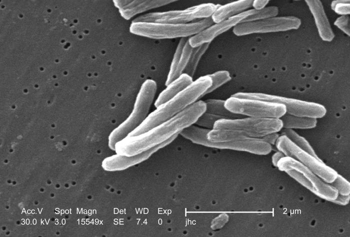
ID#: 8438 Description: Under a high magnification of 15549x, this scanning electron micrograph (SEM) depicted some of the ultrastructural details seen in the cell wall configuration of a number of Gram-positive Mycobacterium tuberculosis bacteria. As an obligate aerobic organism M. tuberculosis can only survive in an environment containing oxygen. This bacterium ranges in length between 2 - 4 microns, and a width between 0.2 - 0.5 microns. TB bacteria become active, and begin to multiply, if the immune system can't stop them from growing. The bacteria attack the body and destroy tissue. If in the lungs, the bacteria can actually create a hole in the lung tissue. Some people develop active TB disease soon after becoming infected, before their immune system can fight off the bacteria. Other people may get sick later, when their immune system becomes weak for another reason. Babies and young children often have weak immune systems. People infected with HIV, the virus that causes AIDS, have very weak immune systems. Other people can have weak immune systems, too, especially people with any of these conditions: substance abuse; diabetes mellitus; silicosis; cancer of the head or neck; leukemia or Hodgkin's disease; severe kidney disease; low body weight; certain medical treatments (such as corticosteroid treatment or organ transplants); specialized treatment for rheumatoid arthritis, or Crohn's disease High Resolution: Right click here and select "Save Target As..." for hi-resolution image (5.48 MB) Content Providers(s): CDC/ Dr. Ray Butler; Janice Carr Provider Email: Creation Date: 2006 Photo Credit: Janice Carr Copyright Restrictions: None - This image is in the public domain and thus free of any copyright restrictions. As a matter of courtesy we request that the content provider be credited and notified in any public or private usage of this image. |
|||
|---|---|---|---|---|
| Source | ||||
| Date | ||||
| Author | ||||
| Permission (Reusing this image) |
|

Historique du fichier
Cliquer sur une date et une heure pour voir le fichier tel qu’il était à ce moment-là
| Date et heure | Dimensions | Utilisateur | Commentaire | |
|---|---|---|---|---|
| actuel | 18 avril 2006 à 21:45 | 700×475 (49 Kio) | Patho | ({{Information| |Description= ID#: 8438 Description: Under a high magnification of 15549x, this scanning electron micrograph (SEM) depicted some of the ultrastructural details seen in the cell wall configuration of a number of Gram-positive Mycobacterium t) |
Pages contenant l’image
La page ci-dessous contient cette image :

