Image:FIPCytology2.jpg
Un article de Wikipédia, l'encyclopédie libre.
Pas de plus haute résolution disponible.
FIPCytology2.jpg (350 × 234 pixels, taille du fichier : 40 Kio, type MIME : image/jpeg)
| | Ce fichier provient de Wikimedia Commons?. Les informations le concernant sont affichées ci-dessous (procédure). |
| Description |
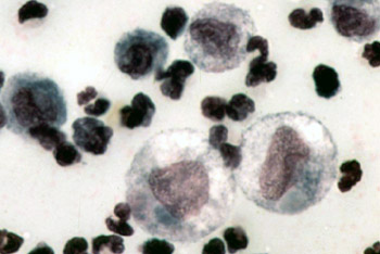
English: Color micrograph of the cytology of FIP-induced effusion. Magnification not specified; estimated to be 1000x.
Original caption: "The cytology of FIP effusion usually contains neutrophils, macrophages and lymphocytes." Image from "Feline Infectious Peritonitis: An Overview of Disease Transmission, Pathogenesis, Signs and Treatment With Emphasis on Diagnosis" ([1]) Clinical Pathology Clerkship Program |
|---|---|
| Source |
Transfered from en.wikipedia |
| Date |
2005-09-30 (original upload date) |
| Author |
Original uploader was Bk0 at en.wikipedia |
| Permission (Reusing this image) |
ATTRIBUTION. |
[edit] License information
 |
The copyright holder of this file allows anyone to use it for any purpose, provided that the copyright holder is properly attributed. Redistribution, derivative work, commercial use, and all other use is permitted.
Aragonés | العربية | Български | Català | Dansk | Deutsch | Ελληνικά | English | Español | Español | Français | עברית | Magyar | Galego | Italiano | 日本語 | 한국어 | Kurdî / كوردی | Latviešu | Nederlands | Norsk (bokmål) | Polski | Português | Svenska | Türkçe | Русский | 中文(简体) | 中文(繁體) | +/- |
cellspacing="8" cellpadding="0" style="width:100%; clear:both; margin:0.5em auto; background-color:#e2f2d2; border:2px solid #acce79;"
[edit] Original upload log
The original description page is/was here. All following user names refer to en.wikipedia.
- 2005-09-30 00:14 Bk0 350×234×8 (40687 bytes) Color micrograph of the cytology of [[Feline infectious peritonitis|FIP]]-induced effusion. Magnification not specified; estimated to be 1000x. Original caption: "The cytology of FIP effusion usually contains neutrophils, macrophages and lymphocytes." I
Historique du fichier
Cliquer sur une date et une heure pour voir le fichier tel qu’il était à ce moment-là
| Date et heure | Dimensions | Utilisateur | Commentaire | |
|---|---|---|---|---|
| actuel | 29 décembre 2007 à 20:53 | 350×234 (40 Kio) | Euthygenes | ({{Information |Description={{en|Color micrograph of the cytology of FIP-induced effusion. Magnification not specified; estimated to be 1000x. Original caption: "The cytology of FIP effusion usually contains neutrophi) |
Pages contenant l’image
La page ci-dessous contient cette image :
Métadonnées
Ce fichier contient des informations supplémentaires probablement ajoutées par l’appareil photo numérique ou le numériseur qui l’a acquis. Si le fichier a été modifié depuis son état original, certains détails peuvent ne pas refléter entièrement l’image modifiée.
| Orientation | Normale |
|---|---|
| Résolution horizontale | 1350 dpi |
| Résolution verticale | 1350 dpi |
| Logiciel utilisé | Adobe Photoshop 7.0 |
| Date de modification | 29 janvier 2004 à 12:43 |
| Espace colorimétrique | 65535 |

