Image:Coarctation and PDA.png
Un article de Wikipédia, l'encyclopédie libre.
Pas de plus haute résolution disponible.
Coarctation_and_PDA.png (555 × 568 pixels, taille du fichier : 43 Kio, type MIME : image/png)
| | Ce fichier provient de Wikimedia Commons?. Les informations le concernant sont affichées ci-dessous (procédure). |
[edit] Summary
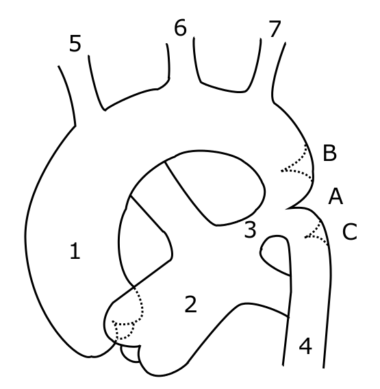
Schematic drawing of alternative locations of a coarctation of the aorta. I, Kjetil Lenes, have made the drawing myself, after information from Valdes-Cruz LM, Cayre RO: Echocardiographic diagnosis of congenital heart disease. Philadelhia, 1998.. Legend: A: ductal coarctation, B: preductal coarctation, C: postductal coarctation. 1: Aorta ascendens, 2: Arteria pulmonalis, 3: Ductus arteriosus, 4: Aorta descendens, 5: Trunchus brachiocephalicus, 6: Arteria carotis communis sinister, 7: Arteria subclavia sinister
The picture is somewhat misleading, with left pulmonary artery crossing behind aorta. This will be changed in a future drawing.
[edit] Licensing
| I, the copyright holder of this work, hereby release it into the public domain. This applies worldwide. In case this is not legally possible: Afrikaans | Alemannisch | Aragonés | العربية | Asturianu | Български | Català | Česky | Cymraeg | Dansk | Deutsch | Eʋegbe | Ελληνικά | English | Español | Esperanto | Euskara | Estremeñu | فارسی | Français | Galego | 한국어 | हिन्दी | Hrvatski | Ido | Bahasa Indonesia | Íslenska | Italiano | עברית | Kurdî / كوردی | Latina | Lietuvių | Latviešu | Magyar | Македонски | Bahasa Melayu | Nederlands | Norsk (bokmål) | Norsk (nynorsk) | 日本語 | Polski | Português | Ripoarisch | Română | Русский | Shqip | Slovenčina | Slovenščina | Српски / Srpski | Svenska | ไทย | Tagalog | Türkçe | Українська | Tiếng Việt | Walon | 中文(简体) | 中文(繁體) | zh-yue-hant | +/- |
Historique du fichier
Cliquer sur une date et une heure pour voir le fichier tel qu’il était à ce moment-là
| Date et heure | Dimensions | Utilisateur | Commentaire | |
|---|---|---|---|---|
| actuel | 23 mai 2006 à 10:14 | 555×568 (43 Kio) | Ekko | (Schematic drawing of alternative locations of a coarctation of the aorta. I, Kjetil Lenes, have made the drawing myself, after information from Valdes-Cruz LM, Cayre RO: ''Echocardiographic diagnosis of congenital heart disease.'' Philadelhia, 1998.. Lege) |
Pages contenant l’image
La page ci-dessous contient cette image :

