Image:Right bundle branch block ECG characteristics.png
Un article de Wikipédia, l'encyclopédie libre.
Pas de plus haute résolution disponible.
Right_bundle_branch_block_ECG_characteristics.png (218 × 200 pixels, taille du fichier : 10 Kio, type MIME : image/png)
| | Ce fichier provient de Wikimedia Commons?. Les informations le concernant sont affichées ci-dessous (procédure). |
[edit] Summary
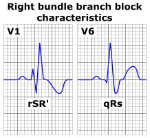
- Description: The characteristic wave patterns of a typical right bundle branch block as seen in an ECG. Only the precordial lead V1 and V6 are shown. Wide QRS complexes are present and there's T wave inversion in lead V1 which is normal in this condition. Note the typical wide and deep s wave in V6. The small q wave in V6 may not always be present. Below each QRS complex is its designation (rSR' and qRs) according to nomenclature.
- Source: I drew this image in Xara X¹ using my own knowledge and several sources for checking whether I drew the image correctly.
- Date: 2 January 2006
- Author: A. Rad
- Permission: See licensing info: GFDL-self
- Other versions: If you require other versions (or have other comments), contact me on my en:Wikipedia user talk page.
- See also "Left bundle branch block ECG characteristics.png" on Wikipedia.
Unfortunately, XaraX¹ can not export to svg.
[edit] Licensing

Historique du fichier
Cliquer sur une date et une heure pour voir le fichier tel qu’il était à ce moment-là
| Date et heure | Dimensions | Utilisateur | Commentaire | |
|---|---|---|---|---|
| actuel | 2 janvier 2006 à 19:18 | 218×200 (10 Kio) | A. Rad | (*Typical right bundle branch block ECG characteristics. *Author & date: —~~~~ *Drawn in XaraX¹ (sorry, no svg export) Category:Physiology Category:Medicine) |
Pages contenant l’image
La page ci-dessous contient cette image :

