Image:Epithelial-cells.jpg
Un article de Wikipédia, l'encyclopédie libre.
Pas de plus haute résolution disponible.
Epithelial-cells.jpg (202 × 202 pixels, taille du fichier : 46 Kio, type MIME : image/jpeg)
| | Ce fichier provient de Wikimedia Commons?. Les informations le concernant sont affichées ci-dessous (procédure). |
-
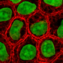
English: Cultured MDCK en:wikipedia:epithelial cells were stained for en:wikipedia:keratin, desmoplakin, and en:wikipedia:DNA. The stained cells were visualized by scanning laser confocal microscopy. The image shows how keratin cytoskeletal filaments are concentrated around the edge of the cells and merge into the desmoplakin which is located at en:wikipedia:desmosomes of the surface membrane. The network of keratin to desmosome to keratin linking the cells of an epithelial sheet is what holds together tissues like skin.
- This image is taken from the wikibooks Cell Biology textbook (licensed under the GFDL): http://wikibooks.org/wiki/Image:Keratin.jpg
- The copyright to this image is retained by John Schmidt (user:JWSchmidt).
- Permission is granted to copy, distribute and/or modify this image under the terms of the GFDL, as indicated in the fine print at the bottom of this page. If you do not want to use this image under the terms of the GFDL, you can alternatively use it under the terms of the Creative Commons Attribution-NonCommercial-ShareAlike License.

This image can be used under the terms of either the GFDL or the Creative Commons Attribution-NonCommercial-ShareAlike License.
Historique du fichier
Cliquer sur une date et une heure pour voir le fichier tel qu’il était à ce moment-là
| Date et heure | Dimensions | Utilisateur | Commentaire | |
|---|---|---|---|---|
| actuel | 2 mai 2005 à 21:49 | 202×202 (46 Kio) | Helix84 | (Cultured MDCK epithelial cells were stained for keratin, desmoplakin, and DNA. The stained cells were visualized by scanning laser confocal microscopy. The image shows how keratin [[Cytoskeleton|cytoskele) |
Pages contenant l’image
Les pages ci-dessous contiennent cette image :

