Image:Blotautoradiogram.png
Un article de Wikipédia, l'encyclopédie libre.
Pas de plus haute résolution disponible.
Blotautoradiogram.png (124 × 105 pixels, taille du fichier : 7 Kio, type MIME : image/png)
| | Ce fichier provient de Wikimedia Commons?. Les informations le concernant sont affichées ci-dessous (procédure). |
[edit] Summary
From enwiki en:Image:Blotautoradiogram.png, uploaded there by en:User:JWSchmidt. The text below from accompanying the image at enwiki
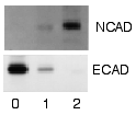
Example of a Western blot using radioactivity. This is an image of a sheet of film that was used to detect radioactively labeled Protein A on a sheet of nitrocellulose. Portions of two different gels (upper and lower panels) were transferred to the nitrocellulose. One was reacted with a primary antibody specific for the cell-to-cell adhesion protein N-cadherin, the other with anti-E-cadherin antibody. The three lanes (0, 1, 2) contained cell proteins from different times (days) after exposure to a chemical inducer of cell differentiation. The results show that when the cells differentiated they shifted from expressing one cadherin to the other.
Source: my personal image.
Uploaded for the Western blot page.
The copyright to this image is retained by John Schmidt (JWSchmidt).
Permission is granted to copy, distribute and/or modify this image under the terms of the Wikipedia GFDL, as indicated in the fine print at the bottom of this page.
If you do not want to use this image under the terms of the GFDL, you can alternatively use it under the terms of the cc-by-nc-sa license.
[edit] Licensing

Historique du fichier
Cliquer sur une date et une heure pour voir le fichier tel qu’il était à ce moment-là
| Date et heure | Dimensions | Utilisateur | Commentaire | |
|---|---|---|---|---|
| actuel | 8 décembre 2005 à 07:22 | 124×105 (7 Kio) | Habj | (From enwiki en:Image:Blotautoradiogram.png, uploaded there by en:User:JWSchmidt. The text below from accompanying the image at enwiki ------ Example of a Western blot using radioactivity. This is an image of a sheet of film that was used to dete) |
Pages contenant l’image
La page ci-dessous contient cette image :

